College of LONG TERM CARE

LOWER EXTREMITY
WOUNDS
If you are a nurse and would like to learn more about wound care and become wound certified, please contact us at(866) 968-6380 to register for a live course or register for our online course at https://www.collegeltc.com/online-course
Dry Eschar

This is a dry eschar of the heel. The eschar acts as a biologic dressing and should not be debrided, unless there is underlying infection as noted by draining pus. Patients who have lower extremity ulcers of any kind should always have peripheral arterial disease, venous insufficiency, and diabetic disease ruled out as a a cause prior to classifying the wound as a pressure ulcer. The appropriate assessment for vascular disease can be seen below. Recommended treatment is to offload the heel with proper devices or pillows. Heel protectors are not a recommended form of offloading. The site should be kept clean, dry, and intact. Certain dressings and ointments can be used on these wounds which include but are not limited to 1. Granulex, 2. Xenaderm, 3. Proderm, 4. Betadiene (controversial), 5. Iodosorb (controversial), or cover with gauze without ointments or creams.
Prevalon Boots

Prevalon boots are appropriate offloading devices for lower extremity wounds, as they remove pressure contact from the surface of the heel and malleolus.
Dry Gangrene

Dry Gangrene of the lower extremity and distal toes must be managed carefully, as it can rapidly decline into wet gangrene, which is life threatening. In general Dry Gangrene is a localized process without evidence of cellulitis, no purulent drainage, no evidence of systemic infection, with fever, or septic symptoms. Dry gangrene can be managed expectantly, but may decline. Wet gangrene must be treated with systemic antibiotics and removal of infection either with amputation or aggressive debridement. Proper treatment for Dry Gangrene is to protect the area from trauma, and can be managed by keeping the area dry and clean. Because the majority of these wounds result from vascular disease, all pressure should be removed with specialized cages for bed sheets, do not use socks, etc.
The Medial Malleolar Artery

Palpation of Medial Malleolus pulse to assess for clinical arterial disease. The medial malleolar artery is located behind the tendons of the extensor hallucis longus and tibialis anterior muscles, to the medial side of the ankle.
After Treatment

Heel protectors are not an appropriate offloading device, as they do not offload the heel from the mattress surface.
Advanced Healing Cases
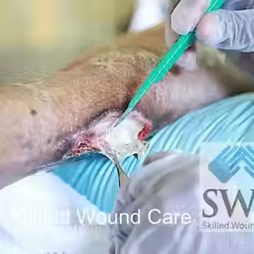
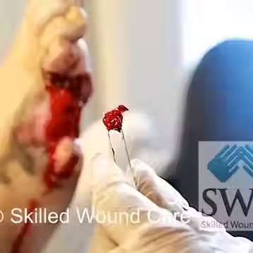
Debridement procedure
Advanced Healing Cases

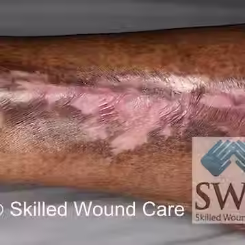
Healing process
Advanced Healing Cases
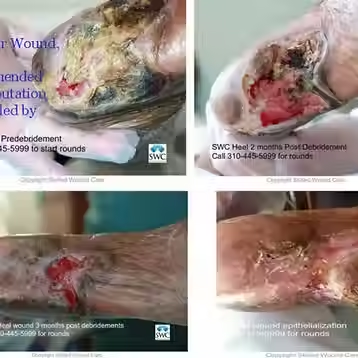
The case above was initially referred for lower extremity amputation. After the family refused amputation and the patient was admitted to a nursing facility, the case was referred to Skilled Wound Care for limb salvage. Vascular work-up demonstrated complete occlusion of the lower extremity arterial supply. The wound was initially debrided to remove infected tissue, not healing. However, after a month of bedside treatment with serial surgical debridement, application of enzymatic debriding agent Santyl, the wound started to heal and develop granulation tissue. After approximately 3 months greater than 80% of the wound epithelialized and the wound eventually re-epithelialized. The case above once again represents healing even in the most challenging arterial wounds, antithetical to what is normally taught in medical texts.
Advanced Healing Cases

The wound on the left is a characteristic Venous Stasis Ulcer (VSU). VSUs are typically wet, draining wounds in the lower leg; versus arterial ulcers which are typically dry wounds. VSUs are usually located on the medial aspect of the leg and malleolus.
Blister

This is a blood filled blister of the heel. Blood blisters are classified as suspected Deep Tissue Injury according to the NPUAP guidelines. In general these should not immediately be classified as pressure ulcers, as they may be caused by underlying vascular or diabetic disease. A proper assessment and work-up must be done prior to diagnosis. These blisters should not be popped, opened, or unroofed. They must be left intact, clean, and dry. Certain dressings and ointments can be used on these wounds which include but are not limited to 1. Granulex, 2. Xenaderm, 3. Proderm, 4. Betadiene (controversial), 5. Iodosorb (controversial), or cover with gauze without ointments or creams.
Wound Care

Great care must be taken when applying wound dressings in the form of kerlix gauze wraps to wound dressings. If the wrapping is too tight it may cause further arterial occlusion and the formation of additional wounds.
Dorsalis Pedis Pulse

Assessment of the dorsalis pedis pulse is an important component in determining the etiology of the lower extremity chronic wound. The dorsalis pedis artery can be palpated or found lateral to the extensor hallucis longus tendon on the dorsal surface of the foot, distal to the navicular bone.
Foot Ulcers

Diabetic foot ulcers are notoriously difficult to heal. Diabetes is a disease which leads to insulin resistance and elevated blood sugars. Over time the disease causes medium and small sized arterial calcification, visual impairment, sensorimotor decline, and impaired immunity. Diabetic ulcers result from trauma in patients who have advanced diabetes. The wounds must be protected and kept clean. Diabetes is the leading cause of amputations in the United States. Poorly controlled blood sugars can lead to further worsening of lower extremity diabetic ulcers. The wound above is a chronic wound with 100% granulation tissue in the wound and a calloused edge. Calloused edges are a result of over growth of keratinocytes, important wound healing cells. These edges must be sharply debrided for ingrowth of new tissue. In addition healthy granulating tissue beds like those above can be treated with hydrogels, collagens, and other dressings. Regranex, Platelet Derived Growth Factor, is an optional FDA approved ointment for healing diabetic ulcers.
Advanced Healing Cases

Before treatment
Advanced Healing Cases

After debridement
Advanced Healing Cases
The wound above was treated by the SWC practice over a year after it was found to have underlying infection and lifting eschar. This PAD ulcer was challenging to heal. The wound was healed over a year with multiple bedside serial surgical debridements, aggressive offloading, and Medihoney as the dressing. Contrary to the standard dogma, vascular ulcers in the lower extremity can heal with aggressive care. SWC has pioneered techniques using advanced dressings and bedside staff education to allow healing in even the most challenging cases.
Advanced Healing Cases
The case above was initially referred for lower extremity amputation. After the family refused amputation and the patient was admitted to a nursing facility, the case was referred to Skilled Wound Care for limb salvage. Vascular work-up demonstrated complete occlusion of the lower extremity arterial supply. The wound was initially debrided to remove infected tissue, not healing. However, after a month of bedside treatment with serial surgical debridement, application of enzymatic debriding agent Santyl, the wound started to heal and develop granulation tissue. After approximately 3 months greater than 80% of the wound epithelialized and the wound eventually re-epithelialized. The case above once again represents healing even in the most challenging arterial wounds, antithetical to what is normally taught in medical texts.
Advanced Healing Cases
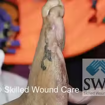
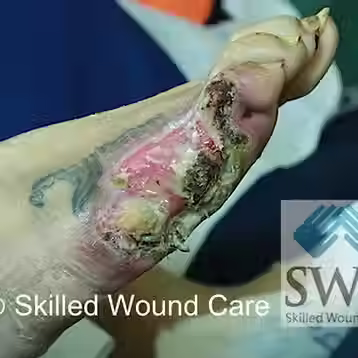
Advanced case of a right lateral foot wound with exposed bone noted with slough and necrosis. It was debrided at bedside in the nursing facility setting. The 5th metatarsal bone was excised to reveal healthy bleeding tissue. Although it has been noted that wound healing cannot occur over exposed bone, Skilled Wound Care has proven the opposite here.
References
-
Peripheral Vascular Disease Author: Everett Stephens, MD, Medscape Reference.
-
Prevalence of peripheral arterial disease in patients at non-high cardiovascular risk. Rationale and design of the PANDORA study Claudio Cimminiello, Claudio Borghi, Serge Kownator, Jean Claude Wautrecht, Christos P Carvounis, Stefanus E Kranendonk, Beat Kindler, Mario Mangrella BMC Cardiovasc Disord. 2010; 10: 35. Published online 2010 August 5. doi: 10.1186/1471-2261-10-35
-
Alzamora et al, Peripheral Arterial Disease Study (PERART): Prevalence and predictive values of asymptomatic peripheral arterial occlusive disease related to cardiovascular morbidity and mortality BMC Public Health 2007, 7:348
-
Joseph E Grey and Keith G Harding. Venous and arterial leg ulcers. BMJ. 2006 February 11; 332(7537): 347–350.
-
The treatment of venous ulcers of the lower extremities. Lonnie L. Whiddon Proc (Bayl Univ Med Cent) 2007 October; 20(4): 363–366.
-
Vincent Rowe. Diabetic Ulcers. Online Medscape Reference.
-
Edwards J et al. Debridement of Diabetic Foot Ulcers. Cochrane Database Syst Rev.2010 Jan 20;(1):CD003556